

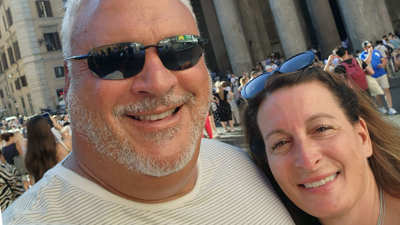
Alex Scherer, a 59-year-old retired Toms River Police Detective, lost both his grandfather and father to heart disease when they were in their 50s. Then in 2024, he lost his sister after she suffered a heart attack. Shortly after, he started noticing some troubling signs himself: He was getting short of breath while working out and climbing stairs, and his ankles were swelling on occasion.
“With all the family history and then noticing some symptoms, I decided to get checked out,” Alex says.
Taking control of his health and getting proactively screened with a new type of imaging technology may have prevented him from having a heart attack.
A New Type of Heart Imaging
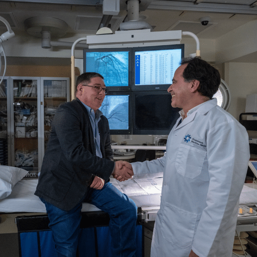
Alex talked about his concerns with a nurse practitioner friend, who referred him to Bharath Sathya, M.D., director of cardiac imaging at Jersey Shore University Medical Center.
Dr. Sathya recommended Alex receive heart imaging from the medical center’s new technology: the Naeotom Alpha.Pro dual-source scanner with photon-counting computed tomography (PCCT). This technology produces images with better resolution in less time compared to traditional computed tomography (CT) scans.
Jersey Shore University Medical Center is among the first hospitals in the U.S. to install this technology, which will only keep increasing in popularity, he predicts. (Hackensack University Medical Center will have the new PCCT later this month; JFK University Medical Center will have it in the future.)
“It was very simple, and it's just like being in an MRI machine. It takes less than 15 minutes. It was no anxiety, no stress,” Alex says.
His results came back showing a 70 percent and a 90 percent blockage in two different arteries, as well as a high calcium score, which indicated his risk of a heart attack was elevated.
“I was shocked,” Alex says. “I always thought because of my family history, maybe I’d have a moderate problem, but I had a severe problem. I felt like a walking time bomb.” He was surprised he’d still been able to exercise and move through his day-to-day responsibilities with such serious blockages.
Two weeks later, he received an outpatient procedure called a coronary angioplasty to open the blocked arteries and place two stents to prevent them from closing again.
An Innovative Approach to Care
To Dr. Sathya, Alex’s experience is a prime example of how PCCT can help people catch heart issues before they become life-threatening. The new technology is part of an ongoing shift in how patients with chest pain and shortness of breath are evaluated, he says.
Previously, evaluations like treadmill stress tests measured the effects on blood flow to identify potential blockages. But these tests don’t actually visualize the structures of the heart that may be blocked. Traditional CT scans produce images of these structures but often overestimate the severity of the issue, leading to a high number of false positives.
PCCT’s more precise image resolution means results are more exact, leading to more accurate diagnoses and timely treatment for patients like Alex, Dr. Sathya says.
Looking Toward the Future
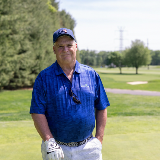
Since his stents were placed, Alex has been able to exercise without getting out of breath, and he’s lost 20 pounds. Under the guidance of his doctors, he’s been adjusting his medication to help prevent further blockages.
“I’m feeling great,” he says. “I’ve been able to get through 30 to 45 minutes of exercise, no issues.”
Dr. Sathya expects more and more patients to be referred for and seek out this kind of imaging. Not only are the results more accurate, but PCCT also delivers a lower dose of radiation than traditional CT scanners, he says. While the benefits of occasional CT scans outweigh the small risk of radiation exposure for most people, they can contribute to cancer risk over time, he notes.
“We really want to make sure patients are being scanned with as little radiation as possible, and doing something with a more advanced scanner helps to achieve that and minimize their risk,” Dr. Sathya says.
It’s important to note that PCCT is not recommended for people with advanced kidney disease. The scan utilizes IV contrast, just like a traditional CT scan, which can put stress on the kidneys.
Alex hopes other people with a family history of heart problems will be proactive about protecting their health. “Get checked out routinely to see if there are any issues,” he says. “Watch your diet and have your blood work done on a consistent basis. I'm very happy that I had this done, and hopefully we took care of the problem, and I can manage it from here on out.”
Next Steps & Resources
- Meet our source(s): Bharath Sathya, M.D.
- Make an appointment online with a cardiology specialist near you, or call 800-822-8905.
- Learn more about heart screenings at Hackensack Meridian Health.
Being at the Right Place at the Right Time Saves Holmdel Man from Cardiac Arrest
When a Holmdel man suffered a severe heart attack, fast action at the hospital gave him a second chance at life.
Cardiac Expertise and Quick Thinking Save Local Mayor’s Heart
When a calcium scan detected arterial blockages, a cardiologist’s quick treatment preserved a patient’s heart.
Man Survives Near Heart Attack Thanks to Valve Replacement
When Gene appeared to be going into heart failure, multiple Hackensack Meridian Health specialists worked together to save his life.
Cardiac Rehabilitation Keeps Brick Man Active After Multiple Heart Surgeries
After multiple heart surgeries, 88-year-old Don Squier proves the power of persistence—logging 1,000 cardiac rehabilitation workouts.