Doctors Explain Fluorescent Guided Brain Surgery

August 26, 2021
By Katie Woehnker
It’s much easier to see where you’re going when your path is well lit, and the same can be said for removing brain tumors.
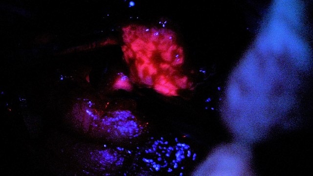
Neurosurgeons are using a new approach where brain tumors glow in the dark, called fluorescent guided surgery. This approach helps surgeons remove malignant brain tumors more aggressively by clearly identifying brain tumor versus healthy tissue.
Neurosurgeon and brain tumor experts William Maggio, M.D. and Aasim Kazmi, M.D. at Jersey Shore University Medical Center, shared how the procedure works and how it’s changed the game for the removal of dangerous brain tumors.
Advancements in Brain Tumor Removal
“Up until now, our eyes and experience have been our main guide in removing brain tumors – discerning what’s normal and abnormal brain tissue,” shares Dr. Maggio.
“Technologies like stealth computer navigation and image guidance use special imaging equipment and computer guidance with a microscope to help in tumor removal, which have helped tremendously, but the fluorescents take it a step further.”
How Does Fluorescent Guided Surgery Work?
A few hours before surgery, the patient will drink a solution – this solution will tend to attach to the tumor tissue in the brain.
“Once in surgery, we’ll have the tumor exposed and use a particular microscope that shines a wavelength of light onto the operating site. This light causes the tumor to light up to a brilliant hot pink color,” explains Dr. Kazmi. “If you look at the brain tissue and tumor under regular light, everything looks normal, but with this microscope we can see exactly where the tumor is.”
The concept is similar to that of a blacklight and a fluorescent poster – as the inks on the poster light up under the black light, the tumor lights up under the microscope light.
Who’s a candidate for this surgery?
“This procedure is typically used for malignant brain tumors, also called glioblastomas,” added Dr. Maggio. “Every patient and case are different, so your surgeon would determine if you’re a candidate for this type of procedure.”
“The goal is to remove only tumor and leave the healthy brain tissue alone. This technique allows us to better see where parts of the tumor are and push the envelope in terms of maximizing the resection,” concludes Dr. Kazmi.
Next Steps & Resources
- Meet Our Experts: William Maggio, M.D., Aasim Kazmi, M.D.
- To make an appointment with Dr. Maggio, Dr. Kazmi or another provider near you, call 800-822-8905 or visit our website.
The material provided through HealthU is intended to be used as general information only and should not replace the advice of your physician. Always consult your physician for individual care.


