
Key Takeaways
- Hackensack Meridian Health is the first hospital network in the Northeast to administer Pixclara for brain cancer imaging.
- Pixclara is an FDA-approved PET imaging drug used with other diagnostic evaluations to help doctors assess recurrent or progressive glioma.
- The imaging option is available for adults and pediatric patients 1 month of age and older.
- Pixclara PET imaging can provide additional information when MRI findings cannot clearly distinguish tumor progression from changes related to treatment.
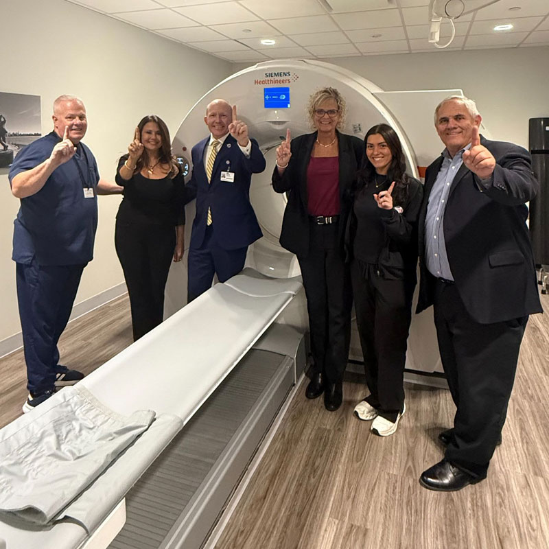
- Hackensack Meridian Health is offering Pixclara at Hackensack Meridian Hoboken Imaging Services.
AI generated.
Hackensack Meridian Health, New Jersey’s largest and most comprehensive health network, announced today that it is the first hospital network in the Northeast United States to administer Pixclara® (floretyrosine F 18), a groundbreaking new diagnostic tool for brain cancer at its ambulatory center at Hackensack Meridian Hoboken Imaging Services.
Recently approved by the U.S. Food and Drug Administration (FDA) and developed by Telix Pharmaceuticals, Pixclara is the first and only positron emission tomography (PET) imaging drug specifically indicated to help physicians differentiate recurrent or progressive glioma from treatment-related changes, in conjunction with other diagnostic evaluations, in adults and pediatric patients 1 month of age and older.
Gliomas are the most common form of central nervous system cancer, making up approximately 80% of all malignant brain tumors, with roughly 24,000 new cases diagnosed in the U.S. each year. Until now, conventional MRI has been the standard of care for imaging these tumors. However, MRIs have inherent limitations, including a lack of biological specificity and an inability to clearly distinguish between actual tumor progression and benign tissue changes caused by radiation or surgery.
“For years, neuro-oncologists have faced the agonizing challenge of looking at an MRI and trying to determine if a brain tumor is growing back, or if the abnormality is simply inflammation and scarring from treatment,” said Dr. Deric Park, Neuro-Oncologist at Hackensack Meridian John Theurer Cancer Center at Hackensack University Medical Center. “Being the first health network in the Northeast to offer Pixclara means our patients now have access to the most advanced diagnostic clarity available for gliomas. It allows our multidisciplinary teams to make faster, more confident, and highly personalized treatment decisions without unnecessary delays.”
When administered intravenously, Pixclara targets specific membrane transport proteins (LAT1 and LAT2) to cross the blood-brain barrier and bind to tumor cells. Once bound, it emits energy that can be detected by a PET scanner. Clinical data shows that combining Pixclara PET imaging (known as FET-PET) with conventional MRI yields a 41% higher diagnostic accuracy than MRI alone.
Because of its high diagnostic accuracy, FET-PET imaging is already recommended in international clinical practice guidelines for the imaging of gliomas, including the NCCN Guidelines®. The FDA’s recent approval of Pixclara finally brings this much-needed technology to the U.S. for both adults and pediatric patients.
“Hackensack Meridian Health is deeply committed to bringing the most innovative, cutting-edge therapies and diagnostics to our communities,” said Mike Horton, Vice President, radiology of Hackensack Meridian Health.