Hackensack University Medical Center Is First in the State and among First Two Centers in the World to Use New Technology to Enhance Assessment of Living Donor Kidneys Before Transplant
March 07, 2022
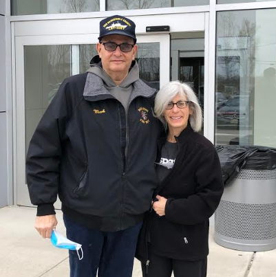
Judy, Kidney Donor & Mark, Kidney Recipient - Both Benefited from IRIS Technology
March is National Kidney Month, a time to raise awareness for the 37 million people in the United States estimated to have chronic kidney disease (CKD). As many as 9 in 10 people are not aware that they have CKD.
Hackensack University Medical Center (HUMC) has become the first center in New Jersey and the second in the world to use an innovative imaging technology before surgery to assess living donor kidneys more precisely and comprehensively than conventional imaging techniques, as well as during surgery to help guide the removal of donor kidneys. HUMC’s living donor kidney surgeons are now employing the advanced imaging software in conjunction with CT scanning to create virtual 3D models of the kidney that assist with preoperative planning as well as intraoperative navigation during robotic-assisted donor nephrectomy
The novel software is called IRIS and is made by Intuitive Surgical, the major manufacturer of robotic surgical systems. The technology has enabled HUMC surgeons to use kidneys that might have been deemed unfit for transplant based solely on the findings of standard CT scanning, potentially expanding the pool of donor kidneys available to patients and freeing them from the need for longer-term dialysis while they wait for a suitable donor kidney.
The virtual anatomical 3D digital models generated by IRIS — which can be downloaded to any iOS device, such as an iPhone or iPad — can be rotated and manipulated by the surgeon to see any part of the kidney in extraordinary detail, including the exact location of arteries, veins, and other structures. Using IRIS can make the removal of a donor kidney faster, more efficient, and safer because the surgeon has more information before and during the surgical procedure.
HUMC urologic surgeons were already very experienced using IRIS to assess the kidneys of patients with kidney cancer before surgery, providing detailed information about a tumor's depth, location, and blood vessels. The technology proved its value for several patients who would have needed the cancerous kidney completely removed if not for IRIS, which provided additional details that enabled patients to have a partial nephrectomy instead (removal of only the tumor while leaving remaining functional kidney tissue in place).
"This technology was so useful for showing that it was feasible to save the kidney in these patients that we began using it to evaluate the kidneys of living donors," explained HUMC’s chief of Living Donor Kidney Surgery and vice chair of Urology, Ravi Munver, MD. Preoperative CT scans often reveal that a donor kidney has abnormal blood vessels or other features that may make it seem unacceptable for transplantation.
In one case, a kidney donor was found to have a tumor, and CT and MRI imaging could not identify the relationship of the tumor to the kidney. IRIS 3D modeling was able to confirm that the tumor not only arose from the kidney, but also that the tumor was not too deep. Dr. Munver performed robotic surgery to remove the kidney, and the tumor was then removed and sent for pathological analysis, as the kidney was placed on a perfusion pump. When pathology showed that the tumor was benign, the kidney was successfully transplanted into the recipient the next day.
Since June 2020, HUMC living donor kidney surgeons have used IRIS technology in more than 50 patients, and this has now become a routine part of their preoperative assessment. "We have not rejected a single donor based on the information provided by this technology, including patients with complex anatomy who might have been rejected based on conventional CT imaging results," asserted Dr. Munver.
One of those donors was Judy Herman, who answered the call for a kidney for Mark Eisen in fall 2020. Judy had known Mark as an acquaintance from the days when she worked as an EMT in a local ambulance corps in Park Ridge, and Mark was a volunteer fireman. In May 2019, Judy had signed up at another hospital to be a living kidney donor and during the assessment, she learned she had a mass on her spleen. The spleen was removed without incident, but when it came time to donate a kidney to Mark, the missing spleen made the operation a bit more complicated. The spleen sits next to the left kidney and acts as a landmark for this organ, but in Judy, the space where the spleen had resided became filled with intestinal tissue.
IRIS technology gave Dr. Munver the information he needed to locate and remove Judy's left kidney laparoscopically which was then transplanted by one of the transplant team surgeons into Mark's body. "IRIS confirmed Judy's anatomy and helped make the surgery more efficient," said Dr. Munver. Judy was able to leave the hospital 24 hours later. Today Mark feels healthier than he has in a long time and considers him and Judy to be "kidney buddies forever."
In fall 2021, Dr. Munver and fellow urologic surgeon Michael Degen, MD, performed the hospital's first robotic surgical donor nephrectomies and used IRIS technology to guide the way. Sitting at the robotic surgical console, the surgeons could plug in an iOS device directly into the robotic system and simultaneously see both the live surgical field as well as the IRIS-generated 3D kidney images, side by side, enabling them to plan the most effective surgical route.
Technologies such as IRIS support HUMC's position as a leading kidney transplant program with some of the best patient outcomes in the region. In the foreseeable future, Dr. Munver and the living donor kidney team have a goal of utilizing IRIS assistance to help remove donor kidneys using single-port robotic surgery, removing the kidney robotically through a single small incision.
Learn more about Hackensack University Medical Center's living donor kidney transplantation program. For an interview contact Mary.McGeever@hmhn.org or call 551-795-1675.