

Radiology and Imaging Services in New Jersey
Advanced Imaging Technology
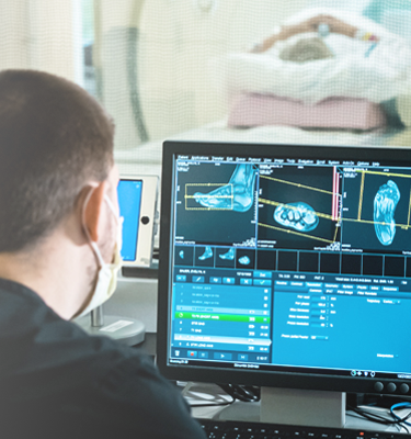
At Hackensack Meridian Health, advanced medical imaging and superior clinical outcomes are driven not only by our state-of-the-art equipment but also by the expertise of our skilled personnel and board-certified physicians. Our radiologists, who are subspecialty-trained in Neuroradiology, Breast Imaging, Musculoskeletal, and Pediatrics, oversee every step of the imaging process, from patient preparation to image acquisition and interpretation.
Our facilities are accredited by the American College of Radiologists, ensuring the highest standards of patient safety and image quality. We offer a comprehensive range of services, including Bone Density, CT, Interventional Radiology, Mammography, MRI, Nuclear Medicine, PET-CT, Ultrasound, and X-ray. With convenient locations throughout New Jersey, we provide a patient-friendly environment with prompt scheduling, flexible hours, and ample parking.
Radiology and Imaging Services We Offer
What is a Bone Density Scan (DEXA Scan)?
A Bone Density Scan, or Dual-Energy X-ray Absorptiometry (DEXA) scan, is a non-invasive imaging technique used to measure bone mineral density (BMD). The procedure uses a low level of X-ray to diagnose osteoporosis, a condition characterized by fragile and brittle bones, and measure bone loss.
Who Should Have a Bone Density Scan?
- Postmenopausal Women: Women are at a higher risk of osteoporosis, especially after menopause due to hormonal changes.
- Elderly Individuals: Aging is a natural factor contributing to bone loss, making older adults more susceptible to osteoporosis.
- Individuals with Risk Factors: Those with a family history of osteoporosis, low body weight, sedentary lifestyle, smoking, excessive alcohol consumption, or long-term use of certain medications (such as corticosteroids) should consider a bone density scan.
- Individuals with Fractures: People who have experienced fractures without significant trauma or have a history of fragility fractures should undergo screening for osteoporosis.
What Can a Bone Density Scan Detect?
- Osteoporosis: DEXA scans can detect low bone mass, a hallmark of osteoporosis, which increases the risk of fractures.
- Osteopenia: This condition refers to bone density that is lower than normal but not low enough to be classified as osteoporosis. It indicates a higher risk of developing osteoporosis in the future.
- Fracture Risk Assessment: By assessing bone density, DEXA scans can estimate the risk of fractures, helping healthcare providers develop preventive strategies.
How to Prepare for a Bone Density Scan
- Clothing: Wear comfortable, loose-fitting clothing without metal buttons or zippers. Avoid garments with metal accessories that may interfere with the scan.
- Medications and Supplements: Inform your healthcare provider about any medications or supplements you are taking, as some may interfere with the accuracy of the scan.
- Avoid Calcium Supplements: Refrain from taking calcium supplements for at least 24 hours before the scan, as they can affect the results.
- Metal Objects: Remove any metal objects, such as jewelry, belts, or eyeglasses, before the scan.
- Inform the Technologist: Inform the technologist if you have had recent imaging procedures involving contrast material or metal implants, as they may affect the scan.
A Bone Density Scan (DEXA Scan) is a safe and painless procedure that plays a crucial role in identifying individuals at risk of fractures and guiding preventive measures. If you fall into any of the aforementioned categories or have concerns about your bone health, consider discussing a bone density scan with one of our providers.
*Not available at Southern Ocean Medical Center.
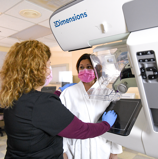
3D Digital Mammography: A 3D mammogram takes multiple x-ray images of the breast and provides a clear and detailed view of breast tissue. The Hackensack Meridian Health network offers the most advanced, high-resolution 3D Mammography technology available , which is supported by Genius AI computer-aided detection. Utilizing the latest AI technology, we offer a breast cancer risk assessment as part of your visit. This enhanced, personalized service helps identify individuals who are at high risk for breast cancer and those who may benefit from genetic counseling.
Breast Health Imaging: Regular screening for breast cancer provides the best chance of finding cancer early when it’s easier to treat and cure. Our Breast Care program offers the most advanced imaging and diagnostic screenings.
Breast Ultrasound: If a suspicious area is detected during a mammogram, or the initial results are not certain, we may recommend a high-resolution ultrasound to provide additional information.
Breast MRI: MRI is highly sensitive imaging that may be used to screen women who have an elevated risk of breast cancer. MRI does not use X-rays.
Cancer Screenings: Regular screenings are an essential part of cancer prevention and provide the best chance of finding cancer early when it’s easier to treat and cure.
Learn more about all of the breast health services we offer.
Cancer Screenings: Regular screenings are an essential part of cancer prevention and provide the best chance of finding cancer early when it’s easier to treat and cure.
Cardiac Computed Tomography Angiography (CCTA): A CCTA is a diagnostic test that produces detailed 3D images of the arteries in the heart to detect abnormalities in how blood flows through the heart and to diagnose cardiovascular disease.
Chest CT Angiogram: An angiogram of the chest uses a special dye and camera (fluoroscopy) to take pictures of the blood flow in the blood vessels in the chest.
Computed Tomography (CT) Scans: CT scans take cross-sectional images of soft tissue or skeletal anatomy inside your body.
Computed Tomography Angiography (CTA): CTA combines a CT scan with an injection of a special dye to create images of blood vessels and tissues in your body.
Photon-counting Computed Tomography (PCCT) Technology: This state-of-the-art system provides diagnostic capabilities across multiple specialties, including Cardiology, Thoracic and Pulmonary Medicine, Neurology, Oncology, and Pediatrics, offering unprecedented image clarity at significantly lower radiation doses.
Contrast Studies: Contrast studies use a special dye to enhance X-ray images of parts of the body that are difficult to view with a conventional x-ray.
Coronary Angiogram: An imaging test that looks at the arteries that supply blood to your heart to diagnose the cause of chest pain or other symptoms. You may also be able to receive treatment during this procedure. (Not offered at Ocean University Medical Center)
CT Calcium Scoring: This scan can help detect calcium-containing plaque in your arteries, which can cause a heart attack.
Head and Neck CT Angiogram: An angiogram of the head and neck uses a special dye and camera (fluoroscopy) to take pictures of the blood flow in the vessels of the head and neck.
Heart Screenings: Heart screenings, prevention tools and diagnostic tests are all services offered by our cardiology specialists to help understand a patient’s heart condition or risk of heart disease, as well as what prevention or treatment options should be used.
Weight-Bearing CT (WBCT): provides full-body, weight-bearing 3D imaging, allowing specialists to see the hips, knees, and feet under natural load.
Angioplasty and Stent Insertion:Angioplasty is a procedure to open narrowed or blocked blood vessels that supply blood to the heart. These blood vessels are called the coronary arteries. A coronary artery stent is a small, metal mesh tube that expands inside a coronary artery. A stent is often placed during or immediately after angioplasty.
Ascitic Tap: An ascitic tap is a medical procedure where a needle is used to drain fluid that is trapped in an internal body cavity, most commonly the abdomen.
Biliary Drainage: Biliary drainage is the insertion of a tube into the bile duct. This is most commonly carried out when the bile ducts are blocked
Bursal Injection: Bursa injections treat bursitis pain. The shot typically contains a steroid like triamcinolone. These anti-inflammatory medicines reduce swelling and pain.
Carotid Stenting: In carotid stenting, a long, hollow tube (catheter) is threaded through the arteries to the narrowed carotid artery in the neck. A metal mesh tube (stent) is inserted into the vessel to serve as a scaffold that helps prevent the artery from narrowing again.
Carpal Tunnel Ultrasound and Injection: If carpal tunnel syndrome is found to be present, ultrasound is used to guide the placement of a needle into the carpal tunnel to inject a small dose of corticosteroid (or 'steroid') and local anesthetic medication.
Image Guided Cervical Nerve Root Sleeve Corticosteroid Injection: A Nerve Root Sleeve Injection is a procedure in which a local anesthetic and steroid solution is injected to relieve pain from irritated and inflamed spinal nerves, which is sometimes caused by the compression of spinal discs.
Image Guided Liver Biopsy: Image guided liver biopsy is a procedure where liver cells are obtained by a needle inserted directly into the liver through the abdominal wall, in the stomach area, and examined.
Image Guided Lumbar Epidural Corticosteroid Injection: An image guided lumbar epidural corticosteroid injection is the accurate placement of a very thin needle, at a given level in this space, under guidance with computed tomography (CT) or X-ray images or pictures to inject corticosteroid (or 'steroid') and usually a long-acting local anesthetic.
Inferior Vena Cava Filters: An inferior vena cava (IVC) filter is a small device that can stop blood clots from going up into the lungs. The inferior vena cava is a large vein in the middle of your body. The device is put in during a short surgery. Veins are the blood vessels that bring oxygen-poor blood and waste products back to the heart.
Joint Injection: A joint injection (intra-articular injection) is a procedure used in the treatment of inflammatory joint conditions. A hypodermic needle is injected into the affected joint where it delivers a dose of any one of many anti-inflammatory agents. The technique may be used to also withdraw excess fluid from the joint.
Nephrostomy: Surgery to make an opening from the outside of the body to the renal pelvis (part of the kidney that collects urine). This may be done to drain urine from a blocked kidney or blocked ureter into a bag outside the body.
Pleural Aspiration: A pleural aspiration is a procedure where a small needle or tube is inserted into the space between the lung and chest wall to remove fluid that has accumulated around the lung.
Radiofrequency Ablation: Radiofrequency ablation (RFA) uses heat to destroy tissue. For pain management, radio waves are sent through a precisely placed needle to heat an area of the nerve. This prevents pain signals from being sent back to your brain. RFA is considered for long-term pain conditions, especially of the neck, lower back or arthritic joints that haven’t been successfully treated with other methods.
SAH Vasospasm Endovascular Treatment: Vasospasm is treated by injecting medication (including verapamil or nimodipine) into the catheter, which is positioned very close to the narrowed artery, to reverse the spasm. Alternatively, a tiny balloon will be inserted through the catheter and into the artery to stretch open the artery (angioplasty).
Selective Internal Radiation Therapy [SIRT]: SIR-Spheres® 0 Selective Internal Radiation Therapy (SIRT) is a treatment for liver cancers or tumors that delivers millions of tiny radioactive microspheres or beads called SIR-Spheres® directly to the liver tumors.
Spinal Cord Embolisation (AVM/DAVF): Spinal Cord DAVF (Dural Arteriovenous Fistula) is an abnormal connection between arteries and veins on the covering (dura) of the spinal cord. DAVF occurs in older people, usually after 50 years of age. DAVF causes abnormal blood flow within the spinal cord and can result in severe spinal disease.
Thyroid fine needle aspiration (FNA): A thyroid fine needle aspiration biopsy is a procedure that removes a small sample of tissue from your thyroid gland. Cells are removed through a small, hollow needle. The sample is sent to the lab for analysis. The thyroid gland is in the front of your neck.
Transarterial Chemoembolization (TACE): Transarterial Chemoembolization, known as TACE, is a minimally-invasive, image-guided treatment for liver cancer. It helps shrink or eradicate tumors by targeting them and blocking their blood flow and delivering chemotherapy directly to the tumor.
Uterine Fibroid Embolisation: Uterine fibroid embolization is a minimally-invasive alternative to hysterectomy or myomectomy. Performed instead of major surgery, this procedure requires minimal or no hospital stay and a shorter recovery. In this procedure, blood supply to the fibroid tumors is blocked, making them shrink.
Varicose Vein Ablation: Venous ablation is an in-office procedure that utilizes radiofrequency energy to cauterize and close bad veins in the legs to alleviate symptoms such as swelling, achiness, fatigue, heaviness of the legs.
Vascular Closure Devices: A vascular closure device is usually a piece of collagen (a fibrous protein found in skin, bone and connective tissue), metallic clip or suture (stitch) designed to provide immediate sealing of the small puncture made in an artery after an angiogram.
Venous Access: Venous access is any method used to access the bloodstream through the veins, either to administer intravenous therapy, parenteral nutrition, to obtain blood for analysis, or to provide an access point for blood-based treatments.
Vertebroplasty: Vertebroplasty is a procedure in which a special cement is injected into a fractured vertebra — with the goal of relieving your spinal pain and restoring your mobility.
Breast MRI: A breast magnetic resonance imaging (MRI) uses magnetic fields to create an image of the breast and is used to screen for breast cancer in women with a higher-than-average risk. Not available at Southern Ocean Medical Center.
Magnetic Resonance Angiography (MRA): MRA uses a magnetic field and radio waves to create a series of thin slice images of arteries and soft tissue in the body.
Magnetic Resonance Imaging (MRI): MRI uses a very powerful magnet and radio waves to diagnose all types of conditions and is especially useful for detecting brain and spinal disorders.
Heart Screenings: Heart screenings, prevention tools and diagnostic tests are all services offered by our cardiology specialists to help understand a patient’s heart condition or risk of heart disease, as well as what prevention or treatment options should be used.
Nuclear Medicine: A type of imaging that uses very small amounts of radioactive material to show your organ structure and function.
Positron emission-computed tomography (PET-CT): PET-CT produces highly sophisticated images that show cell function within the body along with internal anatomy. These are used to diagnose cancer, heart disease, and brain disorders.
Abdominal ultrasound: An abdominal ultrasound is used to assess the liver, gallbladder, pancreas, bile ducts, spleen, and abdominal aorta.
Angioscreen: In just 15 minutes, this simple, non-invasive evaluation will help identify your risk for heart disease and stroke.
Breast Ultrasound: Used to help diagnose an abnormality detected during a physical exam or mammogram.
Diagnostic Ultrasound: Also called sonography, this test uses high-frequency sound waves to produce images of structures within your body.
Elastography: This is a 20 minute Ultrasound of the liver to screen and track progression of Fattty Liver Disease.
Vascular ultrasound: A vascular ultrasound uses soundwaves to see the blood flow in your veins and arteries and detect blockages.
Contrast Studies: Contrast studies use a special dye to enhance X-ray images of parts of the body that are difficult to view with a conventional x-ray.
Fluoroscopy: Fluoroscopy uses contrast dye to view moving parts of the body, producing an “x-ray video.”
X-Ray: An x-ray is a quick and painless test that produces images of the structures inside your body.
Frequently Asked Questions About Imaging Appointments
In most instances a prescription is required. Mammograms do not require a prescription. Please contact the location in advance of your appointment for specifics.
1 Year
X-Rays are walk-in, all other exams require an appointment.
Prescription, ID and insurance card
Generally speaking, wear comfortable clothing that is free of metal.
Yes.
Yes, Hackensack Meridian Health imaging locations are accredited by the American College of Radiology (ACR).
It provides national standards and guidelines for excellence in Radiology.
Results are sent to your referring MD within 24 hours.
Yes, through HMH MyChart
24 hours
Yes.
Patient Resources
What Happens During a Mammogram?
Mammogram preparation and procedure. Learn what to expect, Dr. Graham's advice, and preparation tips. Schedule your mammogram today.

Ultrasound Vs. Mammography
Your doctor may order a breast ultrasound instead of a mammography. What's the difference?

A Lucky Fall
A Lucky Fall: Sea Girt resident Corbett McCarthy's fall led to lifesaving aneurysm detection. Learn how Dr. Sharp's care saved his life.
How to Prep For Your Next Mammogram
Learn how to prepare for a mammogram and what to expect during the procedure. Get tips on what to wear, when to schedule your appointment and more.