

Updated: 5/10/23
Early detection of breast cancer can save lives and increase treatment options that are less disruptive to our daily life. But are all screening methods the same? Harriet Borofsky, M.D., medical director of breast imaging at Bayshore Medical Center and Riverview Medical Center, breaks down the various breast cancer screening technologies and who is best suited for each type.
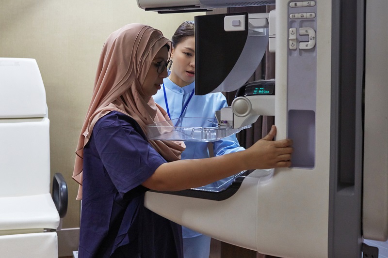
3D Mammography
What is it? 3D-Mammography, also known as Digital Breast Tomosynthesis, is an advancement of Digital Mammography that utilizes multiple exposures as the X-ray tube moves in an arc of the compressed breast. The data acquired is then reformatted by the computer to show thin slices through the breast, improving overall resolution, accuracy and detection, all at the same radiation dose and time.
Does it work? 3D digital mammography not only finds additional cancers, compared to 2D, but also decreases the need for recalls for additional views.
Who is it for? All women who meet criteria for screening and diagnostic imaging. 3D mammograms are now the standard of care and have replaced 2D Digital Mammography.
Contrast Enhanced Mammography
What is it? Contrast enhanced mammography is a special type of mammogram that is performed after injection of a contrast material or dye, to highlight areas of abnormal blood flow and leaky vessels that may indicate an area of concern that otherwise would not be seen on a standard mammogram.
Does it work? By giving contrast, CEM may detect subtle, aggressive tumors hidden on a standard mammogram to increase early detection in certain high risk women improving our ability to find breast cancer as early as possible.
Who is it for? Contrast enhanced mammography may be indicated for women who have an increased risk of breast cancer (a 20 percent to 25 percent and greater chance of getting breast cancer in their lifetime)—including those with gene mutations such as BRCA1 and BRCA2, a family history of breast cancer, certain genetic syndromes or those who had radiation to the chest between age 10 and 30, who are not candidates for MRI. It is an alternative to MRI.
Whole Breast Ultrasound
What is it? Whole breast ultrasound is a non-invasive test that uses sound waves to image the breast tissue. During an ultrasound, the patient lies on an exam table and the technologist places a layer of gel on the breast to scan the entire breast. Whole breast ultrasound does not use ionizing radiation.
Does it work? Ultrasound is typically used as supplemental imaging in women with dense breasts to increase detection of small, early stage tumors. It is also used for additional imaging when something is found on a mammogram or there is an area of clinical concern, such as a lump or focal breast pain. Mammography combined with breast ultrasound may find more breast cancers than mammography alone in women with dense breasts.
Who is it for? Women in the indeterminate risk group (lifetime risk of 12 percent to 20 percent) should get an annual 3D mammogram and strongly consider a supplemental screening test such as whole breast ultrasound. Women in the average risk group with dense breast tissue (a 12 percent or lower chance of getting breast cancer in their lifetime) should receive an annual 3D mammogram and may benefit from supplemental screening tests such as whole breast ultrasound.
MRI (Magnetic Resonance Imaging)
What is it? A breast MRI is an imaging exam that uses a combination of a large magnet, radiofrequency waves and a computer to capture detailed images of the breast after contrast is given by vein. For the procedure, the woman typically lies face down with her breasts positioned through openings in the table. A breast MRI is performed with contrast injected into a vein in the arm during the procedure to make abnormalities clearer. The procedure does not expose patients to radiation.
Does it work? Breast MRI finds significantly more breast cancers that are undetectable in a mammogram or an ultrasound.
Who is it for? Some women at highest risk for breast cancer due to known mutations or with relatives with known mutations, may start screening with MRI at age 25 and mammographic screening by age 30. Women in the high-risk group (20 percent or greater chance of getting breast cancer in their lifetime)—including those with gene mutations such as BRCA1 and BRCA2, a family history of breast cancer, certain genetic syndromes or those who had radiation to the chest between age 10 and 30—should receive an annual mammogram and annual breast MRI with contrast, staggered at 6 month intervals.
Next Steps & Resources:
- Meet our source: Harriet Borofsky, M.D.
- Make an appointment online with a doctor near you, or call 800-822-8905.
- Schedule a breast cancer screening near you.
- CDC: Breast Cancer Risk Factors
Find a doctor near me

Breast Cancer Questions Everyone Should Ask
Every two minutes, a woman in the United States is diagnosed with breast cancer.

What Cancers Can Be Caused By HPV?
Learn about HPV-related cancers. Understand risks & prevention. Protect your health. Schedule a checkup today.

Five Tips for a Healthier Workout
Three of our cardiologists share how to fit heart healthy exercise into even the busiest schedules.

COVID Vaccine and Children: What to Know
Here's everything parents need to know about the COVID vaccine.