Hackensack University Medical Center Lab Creates 3D-Printed Pelvis Model to Help Surgeon Plan Complex Fracture Repair
Physical model informs surgical plan shift from three to two procedures in 17-year-old patient
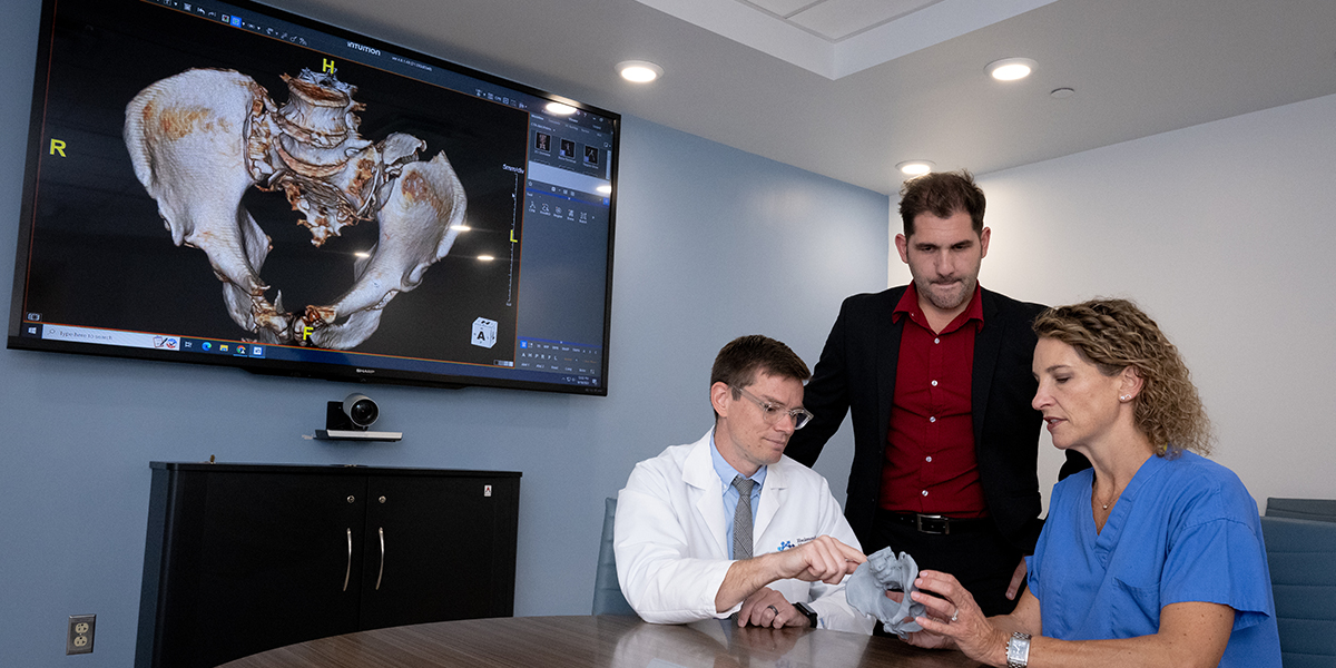
When Hackensack Meridian Hackensack University Medical Center orthopedic trauma surgeon Julie Keller, M.D., called the center’s 3D imaging lab for imaging assistance with a complex pelvic fracture in a 17-year-old patient, lab manager Nick Spagnuolo asked if she’d also like a 3D-printed replica of the injured pelvis.
Her response: “Absolutely!”
Working with the 3D images in this case, Spagnuolo saw the potential for a 3D model to give Dr. Keller additional insight into a complex injury in a young patient. The resulting 3D-printed model gave her perspective that dramatically altered her surgical plan with significant patient benefit.
Collaboration between the orthopedic surgeon, 3D imaging and printing lab and Radiology team brought the model into existence.
To start the process, CT imaging was transferred into modeling software, where Spagnuolo smoothed pixelated surfaces. He worked closely with radiologist Michael McGuire, M.D., to ensure detailed anatomical accuracy in the software map, then initiated printing. This can take a full day as the model design captures detailed anatomy.
The lab can also render and 3D print other anatomy around the affected joint, which can help inform surgical planning in a way in which 3D imaging may be limited. Surgeons can also bring the models into the OR, marked as a cutting guide.
This level of planning and guidance can shave minutes or more off a surgery, or in this case, eliminate an entire procedure. Dr. Keller originally planned a series of three procedures, one from the front and two entering from the back, but the 3D model allowed her to consolidate the repair in two procedures. It also helped her plan the most efficient and effective procedure order.
Dr. Keller could hold the model in her hand and turn it to any angle, where 3D images allow pivots, but have angle limitations. She also found benefit from visually assessing the model at various angles during surgery.
“Especially for a young person we want to give the best possible outcome, and I’m confident we used this tool to give the best possible care,” Dr. Keller said. “This is such great technology and is really on the cutting edge of what is being done around the country.”
The 3D printing lab targets the most complex cases currently, but plans to expand services. In addition to this orthopedic case, they recently collaborated on models for a pediatric cranial case, a pediatric hip repair and a liver tumor, and Dr. Maguire said he sees potential for applications in medical anatomy and surgical training.
Learn more about orthopedic innovations happening at Hackensack University Medical Center.