


Lung Cancer Diagnosis in New Jersey
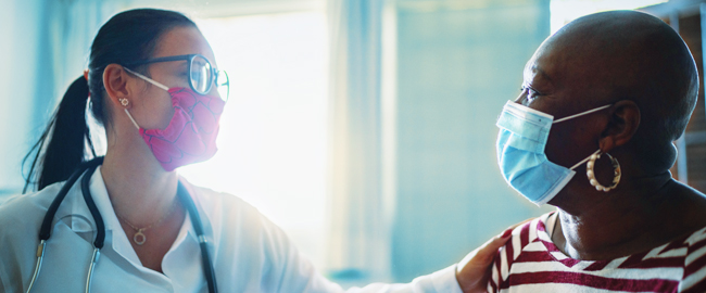
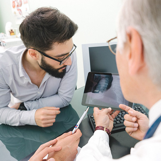
Your doctor may order tests if lung cancer is suspected. We always start with the least invasive way to rule out other conditions. Hackensack Meridian Health offers the most advanced imaging and laboratory testing. Our expert thoracic radiologists and pathologists specialize in identifying and diagnosing lung cancer.
Our Lung Cancer and Thoracic Cancer Specialists
Our lung cancer specialists know the treatment of lung cancers is complex, and we support you every step of the way with comprehensive, customized care.

For More Information
Interested in learning more about Cancer Care services at Hackensack Meridian Health? Click here to request information.
Patient Resources
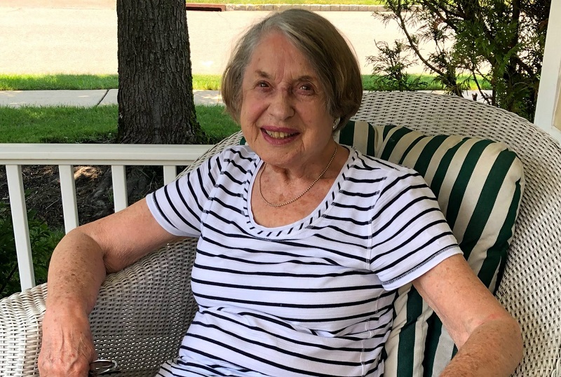
A Fast Recovery from Lung Cancer Surgery
Fast Lung Cancer Surgery Recovery. Learn about minimally invasive options and a patient's positive experience. Call 800-822-8905.
The Facts About Lung Cancer Screening
Learn lung cancer screening facts. Early detection improves survival rates. Simple, low-risk procedure. Call 800-822-8905 to schedule.

The Signs of Vaping Lung Disease
Vaping lung disease symptoms? Learn signs like cough, shortness of breath from Drs. Awad & Awan. Get care at Bayshore Medical Center. Call 800-822-8905.
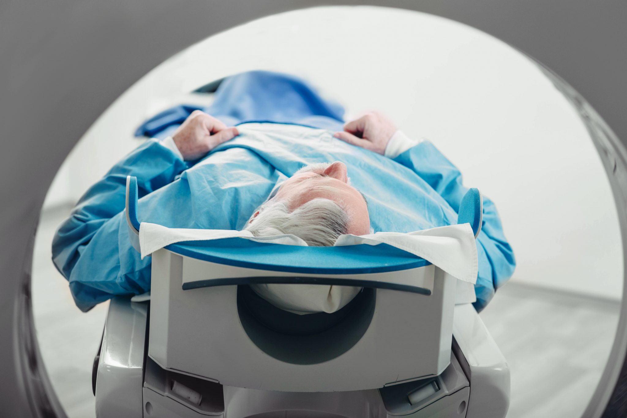
When Should You Get a Lung Cancer Screening?
Lung Cancer Screening: Learn when a low-dose CT scan is right for you. Dr. Rizk explains who should be screened and why. Schedule your appointment today.